
Interventional Oncology
Oncology
Starting from
$3,500
Up to $22,000 depending on centre
Typical duration
3–14 days
Including pre-operative work-up
Content reviewed for medical accuracy by: Dr. Rodina Ainasoa · Medical Content Writer & Health Communications
Overview
A broad, image-guided hub of minimally invasive cancer treatments — tumour ablation, embolisation, and image-guided biopsy — delivered by interventional radiologists using X-ray, ultrasound, CT, or MRI guidance across multiple tumour sites including liver, kidney, lung, and bone, as an alternative or complement to open surgery and systemic chemotherapy.
Your case is matched to a centre on the basis of clinical outcome data, not marketing. We coordinate every step — from pre-operative work-up to post-treatment follow-up — with a dedicated care coordinator.
What's included
Interventional Oncology
Coordinated from
or message us on WhatsApp
No commitment required. Your data is never shared with third parties.
What Interventional Oncology Covers
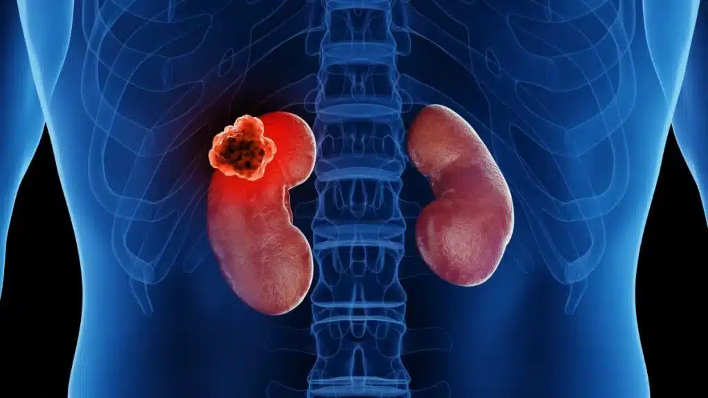
Interventional oncology is a subspecialty of interventional radiology that treats and diagnoses tumors through the skin or blood vessels, guided in real time by X-ray, ultrasound, CT, or MRI — without the open incision of surgery or the whole-body exposure of systemic chemotherapy. It is used for liver, kidney, lung, and bone tumors, either as a primary treatment, a bridge to other therapy, or alongside surgery and systemic therapy, and the technique chosen depends on tumor size, number, and location as assessed by your treating team.
Radiofrequency & Microwave Ablation
A thin probe is guided into the tumor under imaging, then heat generated by radiofrequency current or microwave energy destroys the tumor tissue in place. Commonly used for smaller liver, kidney, and lung tumors.
Cryoablation
Instead of heat, one or more needle-like probes freeze the tumor to destructive temperatures. It is often favored near sensitive structures such as the kidney collecting system or bone, since the ice ball is easier to visualize on imaging during treatment.
Transarterial Embolisation & Chemoembolisation (TACE)
A catheter is threaded through the blood vessels to the artery feeding the tumor, then particles — with or without chemotherapy — are injected to cut off the tumor's blood supply. Most established for liver tumors.
Image-Guided Biopsy
A needle is advanced under CT, ultrasound, or MRI guidance to sample tissue from a suspicious mass, confirming the diagnosis and tumor type before any treatment decision is made.
Palliative Bone Tumor Treatment
Ablation or cement stabilisation (cementoplasty) can relieve pain and reduce fracture risk from bone metastases, often combined with radiotherapy for longer-term control.
The Treatment Process: Step by Step
The general process below applies across tumor sites; your interventional radiologist and the multidisciplinary tumor board confirm which technique applies to your specific tumor after reviewing your imaging and staging.
Imaging review and tumor board discussion
Cross-sectional imaging (CT, MRI, or ultrasound) is reviewed by a multidisciplinary tumor board — including the interventional radiologist, oncologist, and relevant surgeon — to confirm the tumor is a suitable target and to select the technique.
Pre-procedure planning
The interventional radiologist plans the safest needle or catheter path, taking into account the tumor's size and its proximity to blood vessels, nerves, and other organs, and confirms anaesthesia (usually local anaesthesia with sedation, or general anaesthesia for some cases).
Image-guided procedure
Using real-time imaging, the probe, needle, or catheter is advanced through a small skin puncture to the tumor, and the ablation, embolisation, or biopsy is performed percutaneously — without an open surgical incision.
Immediate post-procedure imaging
Imaging is repeated before you leave the procedure suite to confirm the treated area or biopsy site looks as expected and to check for immediate complications such as bleeding.
Recovery & Follow-Up Timeline
Most patients are monitored in a recovery area for a few hours after the procedure to check vital signs and the puncture site before a decision is made on same-day discharge or overnight observation.
The majority of ablation, embolisation, and biopsy procedures are performed as day cases or with a short overnight stay of 24 to 48 hours, considerably shorter than open surgical resection.
Mild pain, low-grade fever, or fatigue (sometimes called post-embolisation syndrome after TACE) can occur in the days following the procedure and typically resolves with simple pain relief within one to two weeks.
Follow-up contrast-enhanced CT or MRI is scheduled to confirm the treated tumor has responded (no residual or recurrent enhancement), with the exact schedule set by your oncology and interventional radiology team.
Source: Society of Interventional Radiology (SIR) and RSNA patient education materials; MD Anderson Cancer Center interventional oncology overview; peer-reviewed interventional oncology literature indexed on PubMed/PMC. Individual timelines vary and are guided by your treating team.
Known Risks & Limitations
- As with any percutaneous or catheter-based procedure, there is a risk of bleeding, infection, or injury to a nearby structure at the needle or catheter entry site or along its path to the tumor.
- Ablation and embolisation are local treatments — they treat the targeted tumor but do not address disease elsewhere in the body, so they are generally combined with surveillance or systemic therapy for tumors that have spread.
- Incomplete ablation, where a rim of tumor at the treated margin is not fully destroyed, is a recognised limitation, particularly for larger tumors, which is why follow-up imaging to confirm complete treatment response is part of standard care.
- Post-embolisation syndrome — transient pain, fever, and fatigue after TACE — is common and expected rather than a sign of failure, but your care team will still monitor you to rule out infection or other complications.
- Not every tumor is suitable for an image-guided approach — size, number, and location relative to major blood vessels, bile ducts, or other organs determine candidacy, so treatment selection is always made by a multidisciplinary tumor board rather than chosen by the patient in advance.
Source: Society of Interventional Radiology (SIR), RSNA, and peer-reviewed interventional oncology outcome studies indexed on PubMed/PMC. No treatment pathway is without risk; your treating team will review your individual risk factors before recommending an approach.
The CureSureMedico Care Pathway
Remote clinical review
Share your recent cross-sectional imaging (CT/MRI/ultrasound), pathology report, and oncology history. Our coordination team forwards your case to an interventional radiologist and the relevant tumor board for initial review before you travel.
Multidisciplinary technique selection
The receiving tumor board reviews your tumor's size, number, and location to confirm whether ablation, embolisation, biopsy, or a combined approach is appropriate, and whether the procedure should be paired with surgery or systemic therapy.
Consolidated treatment plan and estimate
A cost estimate covering consultation, imaging, the recommended procedure, and any inpatient stay is issued before you travel, with the expected timeline and risks of the recommended technique explained clearly.
Procedure and short-stay monitoring
Following the procedure, you are monitored for the expected day-case or short overnight period, and discharged once vital signs and the puncture or access site are stable.
Follow-up imaging and oncology handover
A discharge summary and follow-up imaging schedule is prepared for your home oncology team, since confirming complete tumor response and ongoing surveillance continues well after you return home.
Global cost comparison — Interventional Oncology
Average coordinated costs across our partner centres. Final pricing depends on clinical complexity and chosen hospital.
| Country | Avg. cost | vs. cheapest |
|---|---|---|
🇮🇳 India New Delhi · Chennai · Bengaluru · Hyderabad · Mumbai · Kochi | $4,500 | —chevron_right |
🇹🇷 Turkey Istanbul | $9,000 | +100%chevron_right |
Costs are indicative estimates and vary by procedure specifics, hospital, and clinical complexity. Request a personalised estimate for your specific case.
Frequently asked questions
Not always — it depends on tumour size, number, and location. Interventional oncology techniques such as ablation and embolisation are often used for patients who are not surgical candidates, as a bridge to other treatment, or alongside systemic therapy. Our medical team reviews your imaging and oncology history to determine whether an image-guided approach is appropriate for your tumour type.
No fees. No commitment.
Our guidance is completely free
We are compensated by our partner hospitals — never by patients. You get independent clinical matching, cost transparency, and end-to-end coordination at no cost to you.