
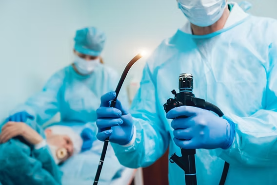
Endoscopic Submucosal Dissection (ESD)
Gastroenterology
Starting from
$3,500
Up to $12,000 depending on centre
Typical duration
5–10 days
Including pre-operative work-up

Content reviewed for medical accuracy by: Dr. Rodina Ainasoa · Medical Content Writer & Health Communications
Overview
An advanced, minimally invasive endoscopic technique for the en-bloc (single-piece) removal of early-stage gastrointestinal cancers and large polyps confined to the mucosa or submucosa — performed entirely through a flexible endoscope passed via the mouth or anus, with no external incisions. Pioneered in Japan and now a standard of care across leading Asian endoscopy centers, ESD avoids open or laparoscopic resection for lesions that meet strict early-stage criteria, preserving the stomach, esophagus, or colon while achieving curative-intent margins.
Your case is matched to a centre on the basis of clinical outcome data, not marketing. We coordinate every step — from pre-operative work-up to post-treatment follow-up — with a dedicated care coordinator.
What's included
Endoscopic Submucosal Dissection (ESD)
Coordinated from
or message us on WhatsApp
No commitment required. Your data is never shared with third parties.
ESD vs. EMR vs. Surgical Resection
Endoscopic submucosal dissection (ESD) removes early-stage GI lesions in one intact piece through a flexible endoscope. It is often compared with endoscopic mucosal resection (EMR), an older piecemeal technique, and with open or laparoscopic surgical resection, which remains necessary for lesions beyond early-stage criteria.
| Parameter | ESD | EMR (Endoscopic Mucosal Resection) | Surgical Resection |
|---|---|---|---|
| What it is | A flexible endoscope with specialised electrosurgical knives is used to dissect beneath a lesion and lift it out in a single intact piece, regardless of size. | A lesion is lifted with a submucosal injection and removed with a snare, typically in fragments (piecemeal) once it exceeds about 2 cm. | Open or laparoscopic removal of part of the stomach, esophagus, or colon, used when a lesion is too deep or too advanced for endoscopic removal. |
| Resection margins & recurrence | En-bloc removal gives pathologists a single specimen, allowing accurate margin assessment and curative-intent resection with a lower local recurrence rate for eligible lesions. | Piecemeal removal makes margins harder to assess and carries a higher local recurrence rate for larger lesions, per ASGE guidance. | Provides definitive margins including lymph node assessment where needed, but at the cost of a more invasive operation. |
| Invasiveness & organ preservation | No external incisions; the stomach, esophagus, or colon is preserved intact aside from the resected lesion. | Also incisionless and organ-preserving, but generally reserved for smaller lesions given its piecemeal limitations. | Removes a larger segment or all of the affected organ in some cases, with longer recovery. |
| Recovery | Typically a short inpatient observation period of one to a few days to monitor for delayed bleeding or perforation, per published ESD case series. | Similar or shorter observation given the smaller lesions typically treated. | Longer hospital stay and recovery consistent with open or laparoscopic abdominal/thoracic surgery. |
| Best suited for | Early-stage lesions confined to the mucosa or submucosa, including larger or flat lesions where en-bloc removal matters most. | Small, straightforward mucosal lesions where piecemeal removal carries acceptable recurrence risk. | Lesions with deeper invasion, lymphovascular involvement, or suspected lymph node spread that fall outside endoscopic curative criteria. |
The Procedure: Step by Step
ESD is performed entirely through a flexible endoscope passed through the mouth or anus, under sedation or general anesthesia depending on the location and complexity of the lesion.
Lesion marking
The endoscopist marks the perimeter of the lesion with cautery dots, guided by prior staging with chromoendoscopy, magnifying endoscopy, or narrow-band imaging.
Submucosal injection
A fluid cushion is injected beneath the lesion to lift it away from the deeper muscle layer, creating a safer working space and reducing perforation risk.
Circumferential incision and submucosal dissection
The endoscopist cuts around the marked lesion and then dissects along the submucosal plane using an electrosurgical knife, freeing the lesion from the underlying wall in one continuous piece.
Specimen retrieval and pathology
The intact (en-bloc) specimen is retrieved and sent for pathology to confirm complete removal with clear margins, which determines whether the resection is curative or further treatment is needed.
Recovery Timeline
Most patients are admitted for observation after the procedure; a large North American series reported that about a third of patients were admitted, with a mean hospital stay of roughly 1.3 days.
Diet is typically restricted or advanced slowly (liquids to soft foods) while the resection site heals, with monitoring for signs of delayed bleeding or perforation.
Most patients return to normal activity and diet within one to two weeks, pending an uncomplicated recovery.
Follow-up endoscopy is scheduled to confirm healing and check for recurrence, with the interval and duration guided by the final pathology result.
Source: American Society for Gastrointestinal Endoscopy (ASGE) guideline on ESD for early esophageal and gastric cancers; published North American ESD case series data. Individual recovery varies and is guided by your care team.
Known Risks & Limitations
- Bleeding — delayed bleeding after ESD has been reported in roughly 2–4% of cases in published series, varying by lesion location.
- Perforation — reported in a similar range, with rates varying by site (higher in the stomach than the esophagus or colorectum in some series); most perforations recognised during the procedure can be closed endoscopically with clips rather than requiring emergency surgery.
- Incomplete or non-curative resection — pathology may show deeper invasion or margin involvement than expected, in which case additional endoscopic treatment or surgery may be recommended.
- ESD requires substantial operator training and experience to achieve high en-bloc resection rates safely; availability and outcomes vary between centres.
- ESD is a flexible-endoscope technique, not a robotic-assisted surgical procedure — it should not be confused with robotic surgical resection, which is a separate, more invasive option reserved for lesions beyond endoscopic curative criteria.
Source: American Society for Gastrointestinal Endoscopy (ASGE); NCBI/StatPearls; published ESD case series on complication rates by anatomic location. No procedure is without risk; your endoscopist will review your individual risk factors.
The CureSureMedico Care Pathway
Remote clinical review
Share your endoscopy, biopsy, and imaging reports. Our coordination team forwards your case to a specialist endoscopist to confirm whether your lesion meets early-stage ESD criteria.
Staging confirmation
Your partner centre reviews or repeats staging — endoscopic ultrasound, chromoendoscopy, or magnifying/narrow-band imaging — to confirm the lesion is confined to the mucosa or submucosa before your estimate is finalised.
Consolidated estimate
A cost estimate covering staging, the ESD procedure, pathology, and inpatient observation is issued before you travel.
Procedure & short-stay recovery
ESD is typically followed by a short inpatient observation period; your care team monitors for delayed bleeding or perforation and reviews the pathology result with you before discharge.
Continuity of care post-return
A discharge summary, pathology report, and recommended surveillance-endoscopy schedule are shared with your home gastroenterologist.
Global cost comparison — Endoscopic Submucosal Dissection (ESD)
Average coordinated costs across our partner centres. Final pricing depends on clinical complexity and chosen hospital.
| Country | Avg. cost | vs. cheapest |
|---|---|---|
🇯🇵 JP | $9,500 | +138% |
🇰🇷 KR | $7,000 | +75% |
🇮🇳 India New Delhi · Chennai · Bengaluru · Hyderabad · Mumbai · Kochi | $4,000 | —chevron_right |
Costs are indicative estimates and vary by procedure specifics, hospital, and clinical complexity. Request a personalised estimate for your specific case.
Frequently asked questions
ESD is suitable for early-stage lesions confined to the mucosa or submucosa without deep invasion, lymphovascular involvement, or evidence of lymph node spread — assessed beforehand with endoscopic ultrasound and imaging. If staging shows deeper invasion or nodal risk, your care team will recommend surgical resection instead, since ESD is curative only within these strict early-stage criteria.
No fees. No commitment.
Our guidance is completely free
We are compensated by our partner hospitals — never by patients. You get independent clinical matching, cost transparency, and end-to-end coordination at no cost to you.