
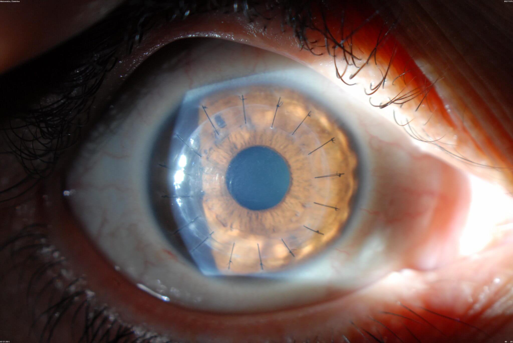
DMEK / DSAEK (Endothelial Keratoplasty)
Ophthalmology
Starting from
$2,200
Up to $9,500 depending on centre
Typical duration
7–14 days
Including pre-operative work-up
Content reviewed for medical accuracy by: Dr. Rodina Ainasoa · Medical Content Writer & Health Communications
Overview
DMEK and DSAEK are selective corneal transplant techniques that replace only the diseased innermost endothelial cell layer of the cornea, leaving the healthy outer layers untouched. They treat corneal endothelial failure — most commonly from Fuchs endothelial corneal dystrophy, bullous keratopathy, or a failed prior corneal graft — and offer faster visual recovery and lower rejection risk than full-thickness corneal transplantation.
Your case is matched to a centre on the basis of clinical outcome data, not marketing. We coordinate every step — from pre-operative work-up to post-treatment follow-up — with a dedicated care coordinator.
What's included
DMEK / DSAEK (Endothelial Keratoplasty)
Coordinated from
or message us on WhatsApp
No commitment required. Your data is never shared with third parties.
DMEK vs. DSAEK: Choosing the Right Endothelial Graft
DMEK and DSAEK both replace only the diseased endothelial cell layer of the cornea, but they differ in graft thickness, surgical handling, and outcomes. Your surgeon will recommend one based on your eye's anatomy and surgical history.
| Parameter | DMEK | DSAEK |
|---|---|---|
| Graft composition | An ultra-thin membrane of Descemet membrane and endothelium only, with no donor stroma, per the American Academy of Ophthalmology (AAO). | A thin layer of donor stroma plus Descemet membrane and endothelium — thicker than a DMEK graft, per the AAO. |
| Surgical handling | The ultra-thin tissue is delicate to unroll and position, requiring advanced technique and a steeper learning curve, per EyeWiki and the Cornea Society. | The added stromal support makes the graft easier to unfold and position, which is why DSAEK is often preferred in more complex or previously operated eyes. |
| Best-corrected visual acuity | Better final visual acuity in comparative studies — one meta-analysis found DMEK achieved a 93% probability of 6/12 vision or better within 5 years, per PMC/NCBI systematic reviews. | Slightly lower final visual acuity than DMEK in the same comparisons — around 83% probability of reaching 6/12 vision within 5 years. |
| Rebubbling rate (graft re-attachment) | A higher rate of partial graft detachment requiring a rebubbling procedure — reported at roughly 18–20% in systematic reviews, per NCBI/PMC. | A somewhat lower rebubbling rate than DMEK in most comparative series, though this varies by study, per NCBI/PMC. |
| 10-year graft rejection risk | The lowest rejection rate among endothelial keratoplasty techniques — around 10% at 10 years in outcomes studies. | A higher rejection rate than DMEK at 10 years — around 19% in the same outcomes studies. |
The Procedure: Step by Step
Both DMEK and DSAEK follow a similar overall sequence, differing mainly in how the donor tissue is prepared and handled.
Pre-operative evaluation
Specular microscopy (endothelial cell count), pachymetry, and slit-lamp examination confirm your candidacy and help your surgeon choose between DMEK and DSAEK.
Donor tissue preparation
Donor tissue is sourced from an accredited eye bank meeting Eye Bank Association of America (EBAA) standards and precision-cut to the required thickness — thinner for DMEK, slightly thicker for DSAEK.
Removal of diseased endothelium & graft insertion
Your damaged endothelial layer is stripped away, and the prepared donor tissue is inserted through a small incision, unrolled, and positioned against the back of your cornea.
Air/gas bubble tamponade
An air or gas bubble is placed inside the eye to press the new graft against the cornea while it attaches naturally over the following hours to days.
Recovery Timeline
You will be positioned to keep the air/gas bubble supporting the graft, and a follow-up exam is scheduled within 24 hours to confirm attachment.
Close monitoring for graft attachment continues; a minority of patients — more often after DMEK — need a rebubbling procedure if the graft has partially detached.
Most patients notice meaningful visual improvement in this window, with steroid eye drops continuing to reduce rejection risk.
Vision continues to improve and stabilise; DMEK tends to reach its best final visual acuity somewhat faster than DSAEK, per comparative outcomes studies.
Source: American Academy of Ophthalmology; Eye Bank Association of America; NCBI/PMC systematic reviews and meta-analyses on DMEK vs. DSAEK outcomes. Individual recovery varies and is guided by your surgical team.
Known Risks & Limitations
- Graft detachment requiring a rebubbling procedure is a recognised part of recovery, occurring more often with DMEK than DSAEK in most comparative series, per NCBI/PMC.
- Graft rejection can occur at any time, though DMEK carries the lowest rejection risk of the endothelial keratoplasty techniques (around 10% at 10 years vs. 19% for DSAEK).
- As with any corneal graft, elevated intraocular pressure (steroid response) can occur with prolonged steroid eye-drop use and is monitored at follow-up visits.
- Primary graft failure — where the donor tissue never attaches or functions properly — is uncommon but can require a repeat procedure.
- As with any procedure involving anaesthesia or sedation, there are anaesthesia-related risks your medical team will review with you individually.
Source: NCBI/PMC systematic reviews on DMEK vs. DSAEK; Eye Bank Association of America. No procedure is without risk; your surgical team will review your individual risk factors.
The CureSureMedico Care Pathway
Remote clinical review
Share your specular microscopy, pachymetry, and diagnosis. Our coordination team forwards your case to a corneal specialist to confirm whether DMEK or DSAEK best suits your eye.
Donor tissue & clinic matching
Your partner centre confirms availability of pre-cut donor endothelial tissue from an accredited eye bank before your estimate is finalised.
Consolidated estimate
A cost estimate covering the procedure, donor tissue, anaesthesia, and initial follow-up (including rebubbling contingency) is issued before you travel.
Surgery & short-stay recovery
Most DMEK/DSAEK procedures are outpatient or short-stay, with slit-lamp follow-up exams scheduled to confirm graft attachment before you are cleared to travel home.
Continuity of care post-return
A treatment summary — including the technique used and a rejection-warning-signs protocol — is shared with your home ophthalmologist for ongoing monitoring.
Global cost comparison — DMEK / DSAEK (Endothelial Keratoplasty)
Average coordinated costs across our partner centres. Final pricing depends on clinical complexity and chosen hospital.
| Country | Avg. cost | vs. cheapest |
|---|---|---|
🇮🇳 India New Delhi · Chennai · Bengaluru · Hyderabad · Mumbai · Kochi | $2,800 | —chevron_right |
🇹🇷 Turkey Istanbul | $5,200 | +86%chevron_right |
🇹🇭 Thailand Bangkok · Phuket · Chiang Mai | $7,800 | +179%chevron_right |
Costs are indicative estimates and vary by procedure specifics, hospital, and clinical complexity. Request a personalised estimate for your specific case.
Frequently asked questions
Both replace only the failing endothelial cell layer, but DMEK transplants an ultra-thin membrane of donor tissue while DSAEK transplants a slightly thicker layer that includes some donor stroma. DMEK tends to deliver better final visual acuity and a lower rejection rate, but the tissue is more delicate to prepare and position, and has a somewhat higher rebubbling rate (needing a small air top-up if the graft partially detaches). DSAEK is technically easier to handle and is often preferred in more complex eyes (e.g. after glaucoma surgery or vitrectomy). Your surgeon will recommend the technique based on your eye's anatomy and surgical history.
No fees. No commitment.
Our guidance is completely free
We are compensated by our partner hospitals — never by patients. You get independent clinical matching, cost transparency, and end-to-end coordination at no cost to you.